De-identified research MRI · Simulated AI outputs · Not for clinical use
Prior
CurrentAnalysed
8annotated change regions across the volume
↗Medical history linked7 dated appointments available
The clinical need · population, problem and outcome
PopulationProblemOutcome
“A way to help neuroradiologists reviewing serial brain MRI examinations overcome acquisition variability and fragmented longitudinal clinical information in order to reduce interpretation time and improve confidence in treatment-response assessment.”
Acquisition variabilityScanner, protocol, orientation and slice differences
Manual comparison burdenRepeated visual review across several timepoints
Fragmented contextImages, reports and treatment history in separate views
Guided product story
Six automated steps. One focused clinical review.
After a study is received from PACS, WAILY prepares the complete longitudinal case in the background. The neuroradiologist enters at step 7 to review, correct and confirm.
The preparation happens before the radiologist reviews.
The following six chapters make WAILY's background processing visible for this concept demo. In clinical use, these steps would run automatically and remain inspectable for traceability.
PACS study received→WAILY prepares the case→Neuroradiologist reviews
01Runs automatically
Load from PACS and clinical systems
Bring the longitudinal case together.
WAILY receives the current and prior FLAIR examinations, previous reports and the relevant dated medical history.
OutputComplete case package2 MRI exams · 7 linked history records
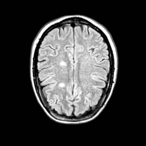
3D FLAIR
421231/47Slice
Prior examination2 Feb 2024 · Reference scan
A
R
L
≈
Current examination16 Feb 2025 · 380 days after reference
A
R
L
Acquisition variation before alignment
02Runs automatically
Registration and normalisation
Align the same anatomy across time.
Orientation, scale and slice position are standardised before any change is measured.
OutputComparable MRI pairAlignment quality 0.92
3D FLAIR
421231/47Slice
Prior examination2 Feb 2024 · Reference scan
A
R
L
Aligned
↔
Current examination16 Feb 2025 · 380 days after reference
A
R
L
Aligned
03Runs automatically
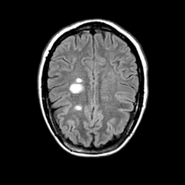
Lesion segmentation
Map candidate lesion regions to their source slices.
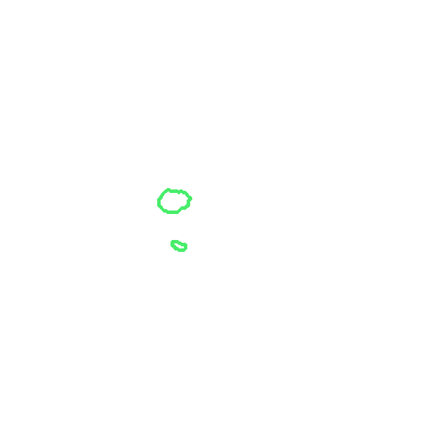
Segmented regions remain directly linked to the underlying MRI so every candidate can be verified.
OutputTraceable lesion contours8 published change regions in this demo
3D FLAIR
421231/47Slice
Prior examination2 Feb 2024 · Reference scan
A
R
L
Registration locked
↔
Current examination16 Feb 2025 · 380 days after reference
A
R
L
Registration locked
Outline key Reference change region Prototype confidence flag, review required
04Runs automatically
Quantitative measurement
Measure lesion burden consistently.
WAILY calculates candidate count, total lesion volume and the largest individual lesion from the segmented examination.
OutputStructured measurementsReady for longitudinal comparison
3D FLAIR
421231/47Slice
Prior examination2 Feb 2024 · Reference scan
A
R
L
Registration locked
↔
Current examination16 Feb 2025 · 380 days after reference
A
R
L
Registration locked
Outline key Reference change region Prototype confidence flag, review required
05Runs automatically
Longitudinal comparison
Separate measurable change from scan variation.
Aligned measurements are compared to surface new, enlarging and stable candidates with visible uncertainty.
OutputChange candidates3 new · 1 enlarging · volume +11.8%
3D FLAIR
421231/47Slice
Prior examination2 Feb 2024 · Reference scan
A
R
L
Registration locked
↔
Current examination16 Feb 2025 · 380 days after reference
A
R
L
Registration locked
Outline key Reference change region Published lesion-change region Prototype confidence flag, review required
06WAILY USP
Longitudinal clinical context
Connect quantitative MRI change with medical history.
Previous reports, diagnoses, medication history and relevant clinical notes are condensed into a dated timeline while the original source remains one click away.
OutputEvidence-linked context7 dated records · sources traceable
Medical history at a glanceSelect any date to open the original record
Selected evidenceCurrent MRI and clinical assessment
Three new and one enlarging lesion candidate were flagged, total volume reached 7.6 mL and recent right-hand paraesthesia was noted.
Interactive longitudinal trendTotal lesion volume over time
Illustrative measurements · days since first recorded visit
16 Feb 20257.6 mL1864 days since first recorded visit
Current MRI and clinical assessment
Three new and one enlarging lesion candidate were flagged, total volume reached 7.6 mL and recent right-hand paraesthesia was noted.
Step 7 · Neuroradiologist interface
The radiologist enters here.
WAILY presents one focused review with aligned images, quantitative change, medical context, uncertainty and links to every supporting source.
Current examination16 Feb 2025 · 380 days after reference
A
R
L
Registration locked
Outline key Reference change region Published lesion-change region Prototype confidence flag, review required
Interactive longitudinal trendTotal lesion volume over time
Illustrative measurements · days since first recorded visit
16 Feb 20257.6 mL1864 days since first recorded visit
Current MRI and clinical assessment
Three new and one enlarging lesion candidate were flagged, total volume reached 7.6 mL and recent right-hand paraesthesia was noted.
Context assistant · WAILY differentiator
Chat with the patient record and MS evidence
One interface for patient-specific questions and general MS imaging knowledge, with the supporting sources kept visible.
Local retrieval concept7 patient recordsCurrent and prior MRICurated MS literature
Suggested questions
Manual review shortcuts
WAILY
Ask about this patient, the longitudinal MRI comparison or general MS imaging knowledge. Each patient-specific answer shows which records support it.
7 patient records2 MRI examinations
Concept simulation. A clinical implementation would use a locally governed, source-linked model. Neuroradiologist confirmation is required.
PatientDEMO-MS-05De-identified research data
Radiologist in control
01 · Case loaded
Bring the longitudinal case together
3D FLAIR
Current and prior brain MRI examinations are loaded with previous reports and relevant medical history.
421231/47Slice
Prior examination2 Feb 2024 · Reference scan
A
R
L
≈
Current examination16 Feb 2025 · 380 days after reference
A
R
L
Acquisition variation before alignment
The complete WAILY workflow
Quantify MRI change, then interpret it with the medical history.
WAILY prepares the longitudinal comparison and brings the relevant clinical evidence into the same review.
01
Align the examinations
Align examinations across scanners, protocols, slice thickness and orientation.
02
Segment and measure
Calculate lesion count, individual volumes and total lesion burden.
03
Quantify longitudinal change
Compare measurements across time and surface new, enlarging and stable lesion candidates.
04 · Core differentiator
Add the medical history
Combine quantitative MRI changes with diagnoses, previous reports, medication and clinical notes. A concise summary supports interpretation while every statement remains linked to its original source.
12-month validation plan
Prove the four risks that matter before full integration.
AlignmentRegistration error and radiologist pass rate
Change detectionNew or enlarging lesion sensitivity and false positives
UncertaintyConfidence calibration and expert agreement
Workflow benefitTime saved and neuroradiologist usability score
Month 0 to 3Data accessSerial MRI, reports and treatment history
Month 3 to 9Build the copilotAlignment, lesion tracking and context layer
Month 9 to 12ValidateRetrospective study and usability testing